Conditions We Treat
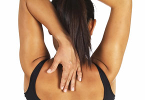
- Back and Neck pain
- Whiplash
- Arthritis
- Post surgery, including joint replacement
- Knee and hip replacement
- General surgery
- All orthopaedic procedures such as arthroscopy, shoulder surgery
- ACL Reconstruction
- All shoulder conditions including frozen shoulder and impingement pain
- Anterior knee pain
- Osteoporosis
- Postural dysfunction and ergonomic advice
- Biomechanical assessment
- Prevention of falls or general decline in mobility
- Soft tissue injuries / sprains and strains
- Fractures
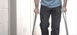
- Mobility issues such as assessment for walking aids or teaching patients how to use crutches on the stairs and regaining confidence going outdoors.
Back pain including prolapsed disc and sciatica.
Back pain is an extremely common problem and affects most people at some point in their life.
It is often triggered by poor posture while sitting or standing, bending, twisting, lifting incorrectly, lack of exercise or weight gain. Anxiety and stress can make your pain worse due to an increase in your muscle tone.
Back ache is most common in the lower back although it can be felt anywhere along the spine.
It is often caused by muscle, tendon or ligament strains but can also be caused by wear and tear (spondylosis) , a bulging or prolapsed disc, sometimes resulting in sciatica (referred leg pain) or other rheumatological conditions such as Ankylosing Spondylitis (where the spine becomes stiff and sore) or Osteoporosis (thinning and weakening of the bones).
During your initial assessment you will be asked specific questions to screen for any serious underlying causes of your back pain. If there are any reasons for concern you will be referred to a doctor or specialist for further investigations, however this is very rare.
Most back pains, including prolapsed discs, get better after a few weeks or months and physiotherapy works exceptionally well to help manage back pain and speed up recovery. Physiotherapy can also help to prevent further episodes of back pain and we will teach you how to adopt a good posture with preventative advice and exercises.
You may be advised to see your GP for analgesia/anti inflammatories/muscle relaxants in the early stages to help alleviate the pain.
A prolapsed (slipped) disc or herniated (bulging) disc occurs when one of the discs that sits between the bones in the spine (vertebrae) is damaged.
In between each vertebra is a protective pad of cartilage called a disc.
It acts as a shock absorber and aids your backs flexibility. The disc has a tough fibrous outer layer (annulus fibrosus) which surrounds an inner gel-like centre (nucleus pulposus).
A prolapsed disc occurs when the outer fibrous layer splits, resulting in the inner 'gel' bulging out of the disc.
As the spinal cord and nerves lie close to the disc, the damaged disc can put pressure on the spinal cord or more commonly, the single nerve root (where the nerve leaves the spinal cord).
As the disc presses on the nerve it causes inflammation, pain and sometimes pins and needles in the area of the nerve and the area of the body controlled by that nerve eg. if it presses on the sciatic nerve at the level of the lower lumbar vertebrae symptoms may be felt in the buttock, thigh, leg and foot.
It can take approximately 6 weeks (or slightly longer) to recover from a prolapsed disc and surgery is only required if the nerve is compressing the spinal cord or nerves which control the bladder/bowel, if there is significant weakness in the leg/foot or if the pain fails to resolve.
In most cases the bulging prolapsed portion of the disc will get smaller and pressure will be released from the nerve.
Physiotherapy can help to:-
- relieve muscle spasm and pain
- reduce inflammation
- mobilise the spinal nerves, joints and soft tissues
- strengthen core muscles
- regain spinal movements
- adopt good posture whilst sitting, standing and walking
- prevent future episodes of back pain.
For more information or to book an appointment call: 07976 980 588 or email physiorehab2u@gmail.com
Whiplash
Whiplash is a term which refers to a neck injury caused by a sudden movement in any direction. Road traffic accidents are the main cause of whiplash although it can be caused from a fall or slip where the head is jolted backwards or a sudden blow to the head, for example during sports, such as rugby.
During a sudden impact the vigorous movement of the head results in over stretching of the tendons, ligaments and muscles in the neck. This can lead to significant stiffness, painful and reduced neck movements, muscle spasms, headaches and occasionally pins and needles into the arm.
Symptoms may not be felt at the time of impact but may develop a few hours later and continue to get worse over the next day or so.
Your physiotherapist will assess you thoroughly and a neurological examination is completed to make sure that there are no serious problems relating to your whiplash. X-ray and scans are rarely required after whiplash injuries and are only carried out if serious pathology such as a fracture is suspected.
Although your neck may be painful it is important to keep it moving from an early stage to improve it's function, prevent muscle wasting (which can lead to instability) and to speed up your recovery. For this reason neck collars are rarely given out today.
Physiotherapists can help you by
- settling the muscle spasm
- reducing the inflammation
- teaching you how to support your neck both during the day and at night by means of postural advice, sitting and sleeping positions.
- Providing a graded exercise programme to keep the neck and nerves mobile to relieve symptoms such as headaches and pins and needles (usually caused by inflammation around the nerve roots in the neck).
- Gentle mobilisations to the spine to release stiff joints following the injury. These are very effective in settling whiplash pain.
The earlier a whiplash is treated the sooner you will recover as the body tends to find alternative ways of moving which can lead to chronic whiplash and faulty movement patterns months down the line from your injury.
Often patients are frightened to move after a whiplash because of the pain and discomfort but your physiotherapist will guide you to make it as comfortable as possible. Often patients require painkillers and anti inflammatories in the early stages to offer pain relief and allow you to move the neck gently.
The time taken to recover from a whiplash varies greatly depending on the severity of the accident.
For more information or to book an appointment call: 07976 980 588 or email physiorehab2u@gmail.com
Arthritis
The two most common types of arthritis are osteoarthritis and rheumatoid arthritis.
Osteoarthritis is the most common type of arthritis and is caused by wear and tear of the joints. There is usually wearing of the cartilage (the smooth lining which covers our bones) and this leads to the formation of bony spurs (osteophytes) or cysts. Eventually the bones rub on one another which causes pain, swelling and stiffness.
Rheumatoid arthritis (RA) occurs when the body's own immune system targets affected joints. This results in painful, swollen, stiff joints.
The tissue that surrounds each joint (synovium) is first affected . This causes inflammation in and around the affected joints. Over time, the inflammation can damage the cartilage, surrounding soft tissues and parts of the bone near to the joint which can lead to joint deformities and instability.
The severity of RA can vary greatly from person to person and it usually has relapses (where the disease flares up) and then periods where it settles down.
Blood tests can be used to help diagnose RA along with x-rays to look for early damage to joints which is characteristic of early RA.
If you are diagnosed with RA you are usually referred to a specialist rheumatology doctor (rheumatologist) so that the correct line of medical treatment can be commenced.
Treatments for RA can include disease-modifying anti rheumatic drugs (DMARDS), biological medicines, Non-steroidal anti-inflammatory drugs (NSAIDs), painkillers and steroids, all of which would be prescribed by a doctor.
Physiotherapy can help to alleviate symptoms associated with both osteoarthritis and rheumatoid arthritis. Gentle exercises can be taught to keep the muscles surrounding the joints as strong and mobile as possible.
If muscles are not used they become weak and this can lead to pain, instability and poor gait patterns.
Physiotherapy can help to calm swollen joints by using treatments such as ice/heat, ultrasound, acupuncture, taping, compressional supports/bracing and walking aids.
Lifestyle advice is usually given to help you to cope with general daily activities.
Surgery is indicated (usually a joint replacement) when the joint damage is so severe that the joint is causing significant pain which affects your daily activities, walking and sleeping.
For more information or to book an appointment call: 07976 980 588 or email physiorehab2u@gmail.com
Post surgery, including joint replacement
A joint replacement may be advised by your orthopaedic consultant if your joint has become very painful, stiff, unstable and other treatments are no longer helping to alleviate your symptoms.
The most common joint replacements carried out are of the knee and hip, although some patients require joint replacement of the shoulder, elbow, ankle or big toe.
During a joint replacement the damaged or diseased part of the joint is removed and a new prosthetic joint is put in it's place. These are made of differing materials depending on the joint being replaced. Sometimes only part of a joint is replaced, especially in younger patients where there is risk of the prosthetic joint wearing out and being replaced again at a later time in life.
There are animated videos available on www.nhs.uk which show how a total hip and knee replacement are carried out (without any gore!).
Physiotherapists ideally like to see their patients who are about to undertake a joint replacement for 'pre-hab' ( pre surgery rehabilitation) so that exercises can be taught and commenced to gain maximum benefit after their surgery. This can be by means of strengthening exercises and stretches to try to gain a better range of joint movement before the new joint is put in place. Patients can be taught how to use crutches for walking and how to tackle stairs or other daily activities such as dressing post surgery.
Post surgery you are sent home from hospital when your surgeon and team are happy that you are safe to do so. The number of days that you stay in hospital are variable depending on the type of surgery, your recovery and pain relief.
Physiotherapy is vital at this time to get the joint moving, and rebuild the strength and mobility of a new joint. Sometimes patients are frightened to move the new joint once home, without the reassurance of a physiotherapist, as there can be some discomfort post surgery. Our domiciliary service enables us to come out to your home and start this vital step to your recovery as soon as possible.
Your physiotherapist will progress you through the varying stages of your recovery, as per the consultants protocol, to help you to regain full mobility, strength and function of the operated joint.
We work very closely with all orthopaedic consultant surgeons so that we work towards the same goal of helping you to recover fully in the least time possible.
We cover all aspects of surgery including general surgery, cardiac surgery and orthopaedic surgery including:
Arthroscopy
A type of keyhole surgery where a camera (arthroscope) is placed inside a joint to help diagnose the cause of the symptoms and often a procedure is carried out to alleviate the symptoms. This may be by shaving off some jagged bone, or removing loose fragments of bone or cartilage.
ACL Reconstruction
The anterior cruciate ligament (ACL) is a tough diagonal ligament inside the knee joint and gives the knee stability. ACL ligament injuries are the most common type of knee injury. It is caused if the lower leg is forced forwards into an extended position or twisted.
If the ACL is torn the knee becomes unstable which can limit movement and function. Balance reactions are also affected and the knee can give way.
Some patients do not require surgery if their knee doesn't feel unstable, there is no pain, no loss of movement and their sport/daily life is not affected.
However with larger tears the ligament may need to be reconstructed by attaching (grafting) new tissue onto it (usually taken from a hamstring or patellar tendon).
Physiotherapy is advised before surgery to:
- Teach exercises to strengthen the knee as much as possible
- Stretch tight tissues to regain the full range of movement in the knee
- Start balance/core training.
This will help with post surgery rehabilitation where your physiotherapist will guide you through your post operative protocol. This is a guided programme of exercises to regain range of movement, strength, function, balance and return to sport or activity.
Many operations are carried out by keyhole surgery due to the benefits of using a minimally invasive technique, however physiotherapy is still required to fully rehabilitate the joint and regain full range, strength, balance and function of the joint affected.
For more information or to book an appointment call: 07976 980 588 or email physiorehab2u@gmail.com
Shoulder conditions
There are many causes of shoulder pain and it is essential that a comprehensive, thorough examination is completed to find out the cause of your shoulder pain.
There are a number of structures that can cause shoulder pain and these include the glenohumeral joint (shoulder ball and socket), the acromioclavicular joint (the joint connecting the collar bone and shoulder blade), the sternoclavicular joint (the joint between the breast bone and the collar bone), scapulothoracic region (area between the shoulder blade and ribs) and the neck.
The two most common shoulder pathologies are frozen shoulder and impingement syndrome.
A frozen shoulder (also known as adhesive capsulitis) is a progressive condition that leads to pain and stiffness in the shoulder. Shoulder movements become reduced and sometimes completely 'frozen'. It is not understood why this condition happens although it is thought that scar tissue forms in the shoulder capsule (tissue which covers and protects the shoulder joint) after a period of inflammation.
Factors that are thought to be associated with frozen shoulder are diabetes, thyroid disease, high cholesterol, heart disease, previous trauma to the affected shoulder and Dupuytrens contracture in the hand. It is more common in women and usually affects people aged between 40-65 years.
Symptoms are pain, then stiffness with increasing limitation of movement which gradually worsens. This can result in muscle wasting around the shoulder as it not used. The affected shoulder becomes painful to lie on at night and daily activities become more difficult (eg reaching up into a cupboard or putting your hand behind your back to dress or reach into a back pocket).
A frozen shoulder can last for approximately 1-3 years however it eventually 'burns out' itself but due to the length of time it can last, most patients seek out treatment to speed up recovery. This can be by means of physiotherapy where exercises are taught to stretch the shoulder capsule, joint mobilisations help to improve the range of shoulder movements and acupuncture can help with pain relief. Sometimes patients are referred to a shoulder specialist for a cortisone injection to aid pain relief. In severe cases patients may require a capsular release, hydrodilatation (where fluid is injected into the shoulder capsule to stretch and break down the scar tissue) or manipulation under anaesthetic.
All of these procedures are carried out by a shoulder specialist surgeon.
Impingement syndrome is another very common shoulder pathology. It is caused by a muscle tendon or bursa (fluid-filled sack) 'catching' in your shoulder as your raise your arm.
The area between the ball of your shoulder joint and the shoulder blade/collar bone is called the sub-acromial space. Your rotator cuff tendons (the 4 rotator cuff tendons attach the muscles from your shoulder blade to the top of your arm and help to stabilise and move the shoulder) run through this space and are cushioned by a fluid-filled sac called the sub-acromial bursa.
If a rotator cuff tendon becomes irritated, inflamed or torn it becomes very painful, especially when your arm is at shoulder level or above. This is because it gets 'trapped' in this position and scrapes against the bone causing pain. Sometimes the tendon develops a build up of calcium deposits, due to long standing inflammation, which are also painful.
Often patients complain of a persistent ache, often felt at the side of the upper arm or pain on overhead movements and also pain at night which disturbs their sleep.
Impingement syndrome is diagnosed by performing specific tests on your shoulder by moving it in a certain way. Often a patient will have a painful arc where pain is felt between 70 - 120 degrees of outwards movement.
Physiotherapy can help to ease symptoms by training the stabilising muscles around the shoulder blade to open the space that the tendon runs through, therefore preventing the tendon from being 'trapped' against the bone. Soft tissue release and stretches help to open up the shoulder joint which again often helps to relieve the pain. Postural and ergonomic advice is given to place the shoulder in the best position whilst at home and work to ease symptoms.
Sometimes painkillers and anti-inflammatory medication are prescribed to relieve symptoms and steroid injections can help reduce the inflammation of the tendons.
If a tear is suspected or your symptoms are not improving you may be referred to a shoulder specialist surgeon for further investigations such as an ultrasound scan or MRI scan. The surgeon will decide if surgery is required and this may be to repair a tear in a rotator cuff tendon or a sub-acromial decompression to widen the space underneath the acromion.
After shoulder surgery you will be referred back for physiotherapy rehabilitation to regain movement and strength in the shoulder and prevent further recurrence of symptoms.
For further information on shoulder conditions and treatment please refer to 'www.shoulderdoc.co.uk'.
For more information or to book an appointment call: 07976 980 588 or email physiorehab2u@gmail.com
Anterior knee pain
Anterior knee pain (patellofemoral pain syndrome) is defined as pain around or behind the knee cap (patella).
Symptoms are usually provoked by squatting, jumping, prolonged sitting with the knees bent or climbing stairs.
Patellofemoral pain syndrome (PFPS) can occur at any age and is a common cause of anterior knee pain in adolescents and young adults. There are many causes of PFPS and these include overuse (eg in sporting activities), malalignment of the patella in the groove on the thigh bone (femur), foot posture (eg flat feet), joint hypermobility or hyperextension of the knees, weakness or tightness of the thigh muscles.
In adolescents, especially those who play sport, Osgood-Schlatter disease is often a cause. This condition affects boys more than girls and usually around the age of 12-15 in boys and 8-12 in girls. It is caused by stresses placed on the tibial tuberosity by contractions from the large thigh muscle (quadriceps) during the adolescent growth spurt.
In middle age and older patellofemoral osteoarthritis (wear and tear) may be a cause.
A biomechanical assessment will be carried out by your physiotherapist and this will help to establish the cause of the pain.
Physiotherapy can help by strengthening weak stabiliser muscles and stretching tight tissues. A foam roller is sometimes advised to help with this. Taping can be very effective and ultrasound is often used to calm inflammation. Sometimes soft knee braces are advised.
You may be advised to buy orthotics to help stabilise the foot to gain better alignment of the patella.
For more information or to book an appointment call: 07976 980 588 or email physiorehab2u@gmail.com
Osteoporosis
Osteoporosis literally means 'porous bones' and it a condition that weakens bones, making them fragile and more likely to break. Our bones are alive and throughout life our bone cells renew to keep our bones strong and healthy. From about the age of 35 we start to lose bone density. This is a normal part of the ageing process, but for some people it can lead to osteoporosis and an increased risk of broken bones (fractures).
Wrist, hip and spinal fractures are the most common type of fractures that affect people with osteoporosis but they can occur in other bones.
It is important that people diagnosed with or at risk of developing osteoporosis take regular exercise, eat healthily, reduce alcohol and stop smoking.
Physiotherapy can be beneficial as
- exercises can be taught to help to strengthen the bones and muscles and prevent tightness around the joints.
- postural advice can be given to help to prevent stooped postures caused by spinal bones altering in shape or following a spinal fracture (the vertebrae become wedged shaped forcing the spine to curve forwards).
- patients can be more susceptible to falls and advice can be given to help reduce the risk by improving balance, gait re-education, issuing a walking aid if this is appropriate.
- breathing exercises can be taught if the shape of the spine has altered to relax muscles and expand the lungs.
- pain relieving techniques can be used to ease any discomfort.
For more information or to book an appointment call: 07976 980 588 or email physiorehab2u@gmail.com
Postural dysfunction
There are a number of different postural types, the two main types being a flat back ( where the lower back loses it's natural curve and has a flat appearance) and a hyper lordotic posture (where the back has an exaggerated lower arch and the bottom tends to stick out).
Other areas of the body adapt to compensate for these postural types and flat backs tend to over curve the middle back (thoracic spine) and round the shoulders. Hyper lordotic postures tend to have tight hips and thighs which is further exaggerated in high heeled shoes.
If your posture is poor it will start to take it's toll and pain can develop in the neck, mid or lower back, hips, knees and can even cause headaches.
Your physiotherapist will complete a full postural assessment (where we assess your body from your head to your toes) and devise a programme of strengthening and stretching exercises to deal with tight or weak muscles.
Core stability exercises and advice will be given to address postures at work, home and for specific activities or sport to help alleviate and prevent further problems relating to your posture.
For more information or to book an appointment call: 07976 980 588 or email physiorehab2u@gmail.com
Biomechanical assessment
A biomechanical assessment is a comprehensive assessment that looks at the structure, alignment and function of your body.
A full assessment from your neck to your feet allows us to find out where there are dysfunctions, weaknesses, instabilities, imbalances and restrictions in joints and soft tissues. These can then be addressed by means of exercises to strengthen, stretch and stabilise. Hands-on treatment to mobilise stiff joints can also help to alleviate the dysfunction or restriction. Postural advice can help rebalance the way in which we move, sit, walk and run.
Poor foot posture can be a major cause of injury and orthotics (insoles worn inside a shoe/trainer) can help to relieve this. Some orthotics can be bought off the shelf but some patients with a more complex foot posture may be referred to a podiatrist for a specialist assessment.
Sports screening is vital for all athletes to help alter any dysfunctions so that unnecessary injuries are prevented.
For more information or to book an appointment call: 07976 980 588 or email physiorehab2u@gmail.com
Falls
1 in 3 people aged over 65 fall every year and this rate increases in the over 85 year olds living in the community. The cost to the NHS is approximately 2.3 billion per year.
The National Institute for Health and Clinical Excellence (NICE) have recommended that older patients with a history of falls should be referred to a physiotherapist to prevent repeated falls.
Physiotherapists can help to prevent falls by:
- Strengthening weak muscles
- Balance training
- Gait re-education
- Assessing the home for hazards which could lead to a fall (eg rugs, wires) -Issuing a walking aid if required -Postural advice as poor posture can affect balance -Stretching tight muscles -Improving confidence and encouraging an active, healthy lifestyle -Promoting independence
A personalised graded exercise programme can be devised, taught and delivered so that patients can practise these safely and independently within their own home.
Soft tissue injuries / sprains and strains
A soft tissue injury (STI) refers to damage of muscles, ligaments and tendons throughout the body.
Common soft tissue injuries usually occur from:
- sprains (an injury to a ligament)
- strains (an injury to a muscle in which the muscle fibres tear as a result of over stretching)
- a direct blow to the muscle resulting in contusion (bruise).
They usually result in swelling, pain, loss of range of movement and function.
Tendonitis refers to inflammation of a tendon and tenosynovitis means inflammation of the tendon sheath that surrounds a tendon. These usually involve an acute injury or several smaller injuries, however if these injuries continue, they can lead to tendon damage and degeneration. In these cases they are referred to as a tendinopathy. A repetitive strain injury (RSI) refers to a soft tissue injury which has been caused by a prolonged or repetitive action. This may be due to activities such as typing, using a mouse, lifting, racket sports such as squash or tennis, writing, gardening, golf, and playing a musical instrument.
Common conditions that arise from tendon inflammation and overuse are tennis elbow (pain on the outer elbow), golfers elbow (pain on the inner elbow), De Quervain's tenosynovitis (pain at the base of the thumb and wrist), carpal tunnel syndrome (pain, weakness and often pins and needles in the palm of the hand) and rotator cuff tendonitis (shoulder pain). Other repetitive actions in the lower limb such as jumping, kneeling, running can lead to tendonitis such as jumpers knee (patella tendinitis over the front of the knee) and Achilles tendonitis (pain and thickening of the Achilles tendon at the back of the heel).
Physiotherapy can help to alleviate these symptoms with many different treatments including soft tissue massage, gentle mobilisations, taping, ultrasound, postural advice and a graded exercise programme dependent on the stage and type of injury.
Fracture
When a patient has a fracture (broken bone) it is usually placed in a cast or splint depending on the type of fracture and the site at which it has occurred. Many patients are given crutches for stable lower limb fractures in A&E departments and then sent home to start their recovery. Some fractures require surgery to realign and stabilise the fracture site.
It is vital that other joints that are not affected by the fracture are kept mobile and strong and physiotherapy can help maintain this. We can also teach you how to get up and down stairs safely with your crutches and teach you how to get on and off the bed or in and out of a chair safely. Many patients are frightened to go outdoors with crutches or walking aids and we can show you how to do this safely and with confidence.
Once you have seen your consultant in fracture clinic and they are happy for you to start moving the area affected, physiotherapists can move the joints within safe limits and progress your rehabilitation programme until you have reached full function again. We can help you to regain a full range of movement, function, strength and return to sport, work or normal activities that you did before the fracture.
Medical Disclaimer
You can read our medical disclaimer here.
For more information or to book an appointment…
Call: 07976 980 588 or email physiorehab2u@gmail.com


